The posterior fornix is the deepest part of the vagina, the pocket that lies behind and below the cervix. It sits directly in front of the pouch of Douglas (the rectouterine cul-de-sac). When the supports here weaken, the top of the vagina widens and organs can begin to descend, so repairs that start at the posterior fornix address laxity where it begins rather than only at the entrance.
Anatomy in plain words
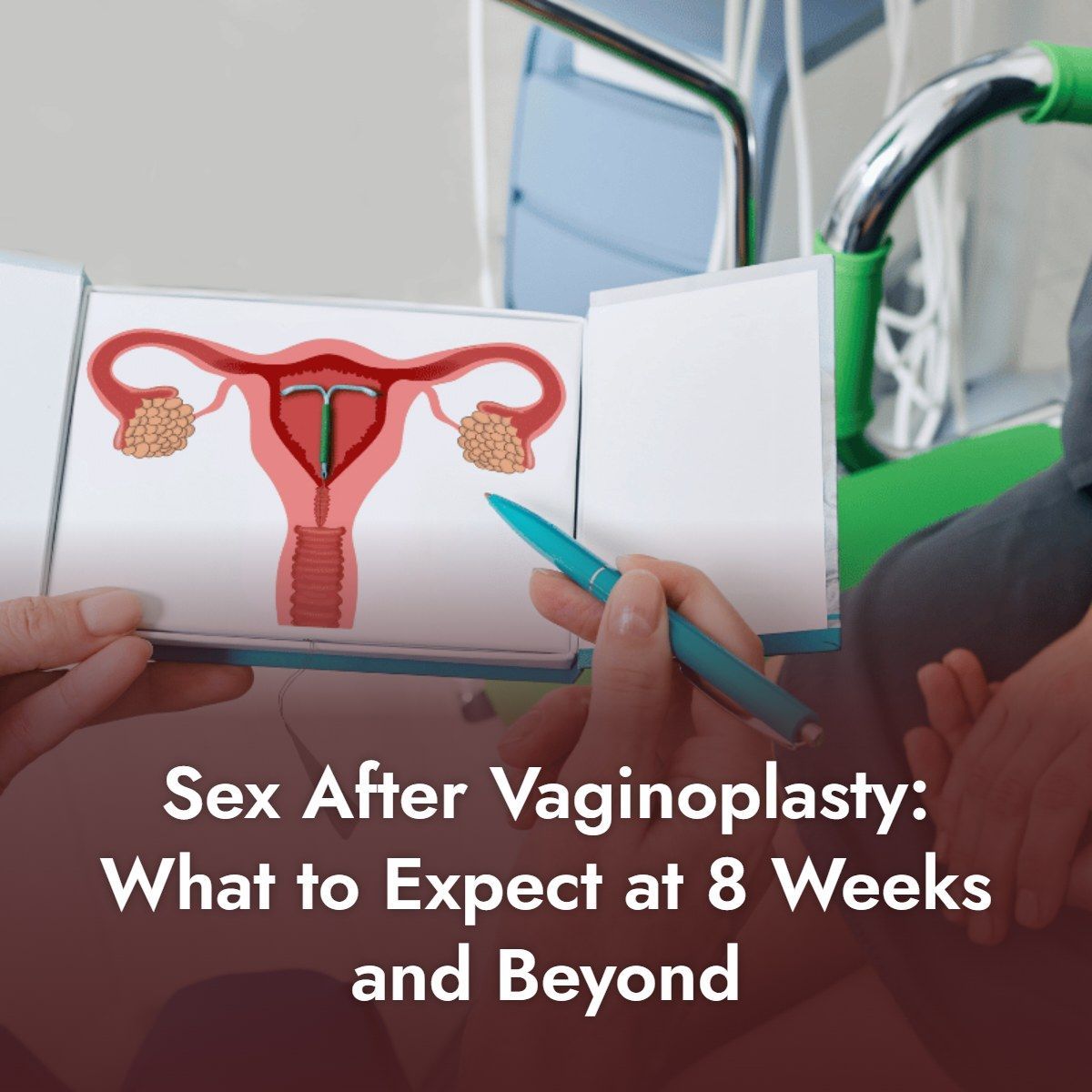
The cervix projects into the upper vagina like a thumb into a glove. The space around it is called the fornix, divided into front (anterior), back (posterior) and side (lateral) parts. The posterior fornix is the largest. Behind its thin wall is the pouch of Douglas, the lowest point of the abdominal cavity, and below that the rectum.
The uterosacral ligaments, which run from the cervix back to the sacrum, hold this region up and back. They are a key part of what gynecologists call Level I support.
What changes after childbirth and with age
- The uterosacral ligaments stretch, so the cervix and fornix sit lower.
- The pouch of Douglas deepens, allowing small bowel to push down (enterocele).
- The back wall below the fornix thins and widens.
- The vaginal axis becomes more vertical, so the canal no longer closes naturally.
Why this matters for tightening procedures
If the deep fornix remains wide and unsupported, tightening the lower vagina narrows only the outlet. Some women then feel a tight entrance with a loose interior, or develop pain from scar at the opening. Repairing from the posterior fornix outward aims to restore the canal along its whole length and to reduce the risk of later prolapse.
What a posterior fornix approach involves
- Assessing cervical position and vaginal length before surgery
- Re-supporting the cervix or vaginal apex using its own ligaments when it has descended
- Closing an enlarged cul-de-sac to prevent bowel from pushing down
- Repairing the back wall and pelvic floor muscle from the fornix to the entrance
- Reconstructing the perineum if needed
Because the fornix lies close to the cul-de-sac and rectum, this work requires experience. Risks include bleeding, infection and, rarely, injury to the bowel.
Other clinical roles of the posterior fornix
The posterior fornix is also where semen pools after intercourse, where a doctor may sample fluid (culdocentesis) and a route for some gynecologic surgery. Its health therefore matters beyond cosmetic concerns.
Signs that the posterior fornix has lost support
Women cannot see the posterior fornix, so changes there are recognized by symptoms and examination rather than by appearance. Common clues include:
- A feeling of looseness deep inside, even though the opening looks normal
- The cervix feeling lower, or being touched more easily during intercourse
- A dragging or heavy sensation that worsens through the day
- Air entering the vagina with position changes
- Low back or pelvic pressure that eases on lying down
- A bulge felt high on the back wall when straining, which may suggest an enterocele
On examination, a gynecologist looks at how far the cervix or vaginal apex descends with straining and how deep the space behind the cervix has become. The POP-Q system records these findings as numerical measurements, which makes it possible to compare before and after treatment.
Not every woman with these symptoms has a problem at the fornix. Similar feelings can come from a weak pelvic floor, a front-wall prolapse or bowel conditions, which is why examination matters more than any single symptom. Ultrasound is sometimes used alongside the physical examination to look at the pelvic floor muscles and organs.
Women who have had a hysterectomy have no cervix, but the same region, now called the vaginal vault, can lose support in a similar way. Symptoms and examination findings are much the same, and repair aims to re-support the vault using the patient's own ligaments where possible.
Deep repair compared with entrance-only and non-surgical options
It helps to understand what each approach can and cannot reach:
- Pelvic floor muscle training strengthens the muscles around the lower vagina. It improves tone and can relieve mild symptoms, but it cannot shorten stretched uterosacral ligaments.
- Energy-based devices such as laser and radiofrequency heat the lining. They may change surface tissue quality but do not re-support the cervix or fornix, and the FDA has warned that their safety and effectiveness for vaginal rejuvenation have not been established.
- Filler and threads add bulk or tension near the entrance and do not repair support; they can cause lumps, infection or migration.
- Entrance-only surgery narrows the opening and perineum. It can help when the problem is limited to Level III but may leave a wide interior.
- Repair starting at the posterior fornix addresses the deep, middle and lower levels together, at the cost of a more involved operation that requires surgical experience.
The right choice depends on where the support has failed. A woman whose cervix and fornix are well supported may need only a lower repair or physiotherapy, while a woman with a deep, wide fornix and early descent of the cervix is more likely to benefit from a repair that starts at the top. Measuring before deciding helps avoid both under-treatment and unnecessary surgery.
It is also reasonable to ask a surgeon how often they perform deep repairs, how they protect the rectum and bowel during dissection, and how they confirm that vaginal length has been preserved at the end of the operation.
Recovery after a repair that begins at the fornix
Although the dissection reaches deeper, recovery is broadly similar to that of other native-tissue vaginal repairs:
- Day 1 to 3: soreness, mostly at the entrance, with light spotting; walking is encouraged
- First week: many women return to desk work; stool softeners help avoid straining
- Weeks 2 to 4: swelling and discharge settle as absorbable sutures dissolve
- Week 6: heavy lifting and exercise usually resume after review
- Weeks 6 to 8: intercourse usually resumes once healing is confirmed
Report heavy bleeding, fever, worsening pain or difficulty passing urine or stool promptly. Avoiding constipation and heavy lifting for the first six weeks protects the deep sutures while they heal.
Most women find that discomfort from the deeper part of the repair is mild, because the upper vagina has fewer pain fibers. A feeling of fullness or pressure is common in the first weeks and usually fades as swelling settles. At the six-week review, the surgeon checks healing, support and vaginal width before clearing intercourse and exercise.
After full recovery, continuing pelvic floor exercises, preventing constipation and avoiding repeated heavy lifting help the deep supports stay in place over the long term.
Women traveling for surgery should plan to stay near the surgeon for at least the first several days, and arrange a check with a local gynecologist if the six-week review cannot be attended in person.
Frequently asked questions
Can I feel my posterior fornix?
Usually as the soft area behind the cervix at the top of the vagina. Its depth varies between women.
Does a hysterectomy remove the fornix?
After hysterectomy, the top of the vagina is closed as the vaginal vault. Supporting that vault remains important to prevent vault prolapse.
Is surgery at the fornix more painful?
The upper vagina has fewer pain fibers than the entrance, so most discomfort after surgery comes from the perineal area.
Is an enterocele dangerous?
An enterocele, where small bowel pushes down behind the vagina, is rarely dangerous but can cause pressure, a bulge and discomfort. It is usually repaired by closing the enlarged cul-de-sac and restoring support at the top of the vagina.
Symptoms often worsen with standing and ease on lying down.
Will a deep repair change vaginal length?
The aim is to keep a normal, functional length. Careful surgeons measure the vagina before and after repair and avoid shortening or narrowing it, which could make intercourse uncomfortable.
If length is a concern for you, ask the surgeon to explain how it will be measured and recorded.
References
- DeLancey JO. Anatomic aspects of vaginal eversion after hysterectomy. American Journal of Obstetrics and Gynecology (1992).
- Barber MD, Maher C. Apical prolapse. International Urogynecology Journal (2013).
- Standring S (ed.). Gray's Anatomy: The Anatomical Basis of Clinical Practice.
This article is for general education and does not replace an examination by a qualified gynecologist. Individual results vary, and every procedure carries risks.